Seven years ago, researchers at Stanford University started an ambitious experiment: They began growing miniature, simplified versions of the human brain from stem cells inside a lab, then later injected that tissue into the brains of newborn rats.
Their results, published Wednesday in the journal Nature, showed that the brain-like human tissue integrated with the rat tissue, then continued to mature.
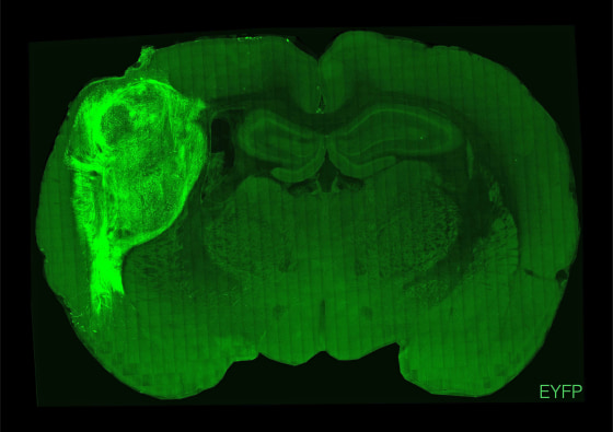
Those brain cells, in turn, seemed to influence the rats’ behavior.
The researchers injected the human tissue into the rats’ somatosensory cortexes — regions that receive and process sensory information like touch or pain. After about two weeks of training, the rats began to lick a spout in search of water whenever the researchers stimulated the human neurons (they did this using blue light lasers). The researchers also used a puff of air to prod the rats’ whiskers, then observed how the human neurons responded.
"We found that human neurons respond very quickly after we stimulated the whiskers. In fact, more than 70% of the human neurons are engaged in some form of activity within a second or so of that stimulation, so that tells us that they’re probably connected," Sergiu Pașca, one of the study’s authors and a professor of psychiatry and behavioral sciences at Stanford, said on a call with reporters.
"Human neurons become part of the rat circuitry," Pașca said, adding that the neurons were "sparkling with electrical activity" under a microscope.
After the transplants, he said, the human neurons grew to six times their original size over about eight months, making up roughly one-third of a single hemisphere in the rat brains.
The rats didn’t show signs of health issues like seizures or epilepsy, which the researchers had worried might arise. More than 70% were alive one year after the transplants.
The study is the latest example of an attempt to transplant human cells into animals. This line of scientific inquiry started decades ago, and some past tries have been successful: In 2006, developmental biologist Ali Brivanlou and a team of researchers at The Rockefeller University showed that they could grow human embryos in mouse tissue. Then in 2013, a group of Belgian researchers transplanted human neurons into newborn mice, creating functional brain circuits.
In 2018, researchers at the Salk Institute for Biological Studies implanted human brain-like structures in mice, producing results similar to the Stanford study. But they used adult mice, whereas the Stanford researchers chose newborn rats in order to see how the human neurons integrated with the rodents’ developing brain circuits.
Brivanlou said the Stanford study is novel because of the way the researchers were able to grow complex, 3D structures that represent the human cortex. The cortex is the most integral part of the brain, where cognitive centers are located.
Growing that type of tissue structure and then transplanting it "has not been done very often; in fact, maybe not at the level of precision that this paper is describing," Brivanlou said.
But novelty is not the end goal. Rather, scientists hope that the ability to perform human-rat transplants will give them a better understanding of how genetic mutations influence brain circuits and affect how people think and behave.
Eventually, this type of rat model could be used to study psychiatric disorders, autism or neurodegenerative diseases like Parkinson’s or Alzheimer’s, and even to identify new treatments or test their effectiveness.
In that sense, the new study could "open the doors to tremendous medical and basic understanding of the way the brain works and, at the same time, what happens when things do not work very well," Brivanlou said.
The Stanford researchers, in fact, used their transplant technique to investigate Timothy syndrome, a rare genetic disorder in humans that can result in life-threatening abnormal heartbeats and may also lead to autism. They transplanted tissues derived from three people with Timothy syndrome into baby rat brains.
They found that those human cells didn’t grow as large inside the rats and weren't as structurally complex as the other human cells. That signaled to the researchers that the genetic mutations responsible for Timothy syndrome in people had impeded the rats’ brain development.
The researchers have yet to study how such mutations change rats’ behavior, however.
Pașca said that although scientists have a lot of information about which genes are linked to psychiatric disorders, they have little understanding of what those genes change inside the brain, or of the nongenetic factors that contribute to neurological disorders. Timothy syndrome is a prime candidate for this type of study, since it’s caused by a mutation in a single gene involved in helping the brain process electrical signals. In the future, rat experiments might help scientists understand more complex neurological conditions.
But Brivanlou acknowledged that this type of research comes with ethical questions.
"How much of a human cell can you put in a mouse embryo for that embryo to still be called a mouse? I think this is a very legitimate question that needs to be addressed," Brivanlou said.
Still, he said, the potential benefits could make the experiments worth pursuing.
“If you have a way to save somebody’s life or cure somebody’s disease, or provide at least some sort of a therapy to minimize the pain or extend the life of the person, you should be allowed to exercise that,” Brivanlou said.
